Cardiovascular Pathology Lab
The Cardiovascular Pathology Laboratory provides innovative, objective pathology support of the utmost quality to improve medical device technologies and subsequently, patients’ lives and creating learning opportunities and new knowledge for students and the scientific community.
Services
- Gross Evaluation:
- The CVP lab can do gross evaluations on a fee for service basis. We specialize in explanting and evaluating cardiovascular devices.
- Gross photography and sectioning:
- On a fee for service basis, gross pathological evaluation of the tissues, organs, and medical device in situ is available by a trained pathologist. We use multiple high-resolution Canon Digital SLR cameras to capture images.
- Histology:
- Exakt grinding and tissue embedding: Using the Exakt grinding and tissue embedding system, slide samples for histology can be prepared for imaging and analysis of metal, bone, and other hard samples. Our trained technicians can produce microscopic sections of sample at less than 20 micrometers thickness and stain with H&E and Trichrome.
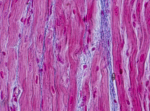
- Paraffin embedding: CVP offers traditional paraffin-embedding histology and routinely performs H&E, Trichrome and PTAH staining.
- Micro-X-Ray and Micro-CT Imaging:
- Our NSI X-50 CT and X-ray images under magnification to look for fractures or other internal structural damage in stents and other medical devices. A movable bar allows the sample to be precisely rotated to observe sample features at any angle under a wide range of magnification.
- The NSI eFX software allows highly detailed CT reconstructions to be analyzed in three dimensions and separated into digital layers by material density or radio-opacity.
- Using this modality, medical devices embedded in tissue can be reconstructed precisely as a 3-D model without removing the device or compromising the tissue.
- Contrast Imaging:
- TAMU CVP also has the capabilities to inject vessels with liquid barium or other contrast agents such as Iohexol. This allows for better evaluation of the vessels.
- Scanning Electron Microscopy Hitachi TM3000 Tabletop SEM:
- This low vacuum SEM, capable of magnifications of 30,000X, will be useful in analyzing specimens before histology is performed.
- The SEM suite includes:
- Motorized stage to allow more control of the images taken to make a map of the surface
- A stent adaptor that will permit visualization of all outer surfaces without removing the specimen from the vacuum chamber.
- 3D view software.
- Energy-dispersive X-ray spectroscopy (EDS) capabilities to determine the surface composition of samples
- Histological Analysis
- Using digital microscopy and powerful image analysis software, stented arterial sections can be evaluated parametrically to determine the level of stenosis and neointimal thickening.
- Qualified pathology staff also provide a quantitative score of the level of damage done to the test artery by evaluating the following: Fibrosis, RBC count, Multi-nucleated giant cells (MNGC), Macrophages.
- Using similar techniques, a pathologist can qualitatively measure inflammation, RBC distribution, etc. by assigning a score to each category.
- Pathology Reports
The CVP Lab offers preliminary reports written by a trained pathologist. After further evaluation (light microscopy, electron microscopy, radiography), the CVP lab will prepare a final report with all findings from evaluation. These reports can include photo montages with gross, microscopic, X-Ray, CT, and SEM/EDS images. After evaluating multiple specimens from the same protocol, the CVP Lab can prepare an executive summary to summarize key findings throughout the entire study.

