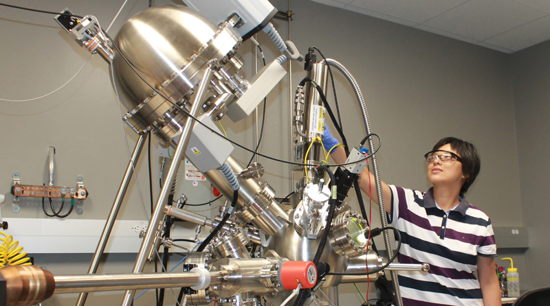
Materials Characterization Facility
The Materials Characterization Facility (MCF) at Texas A&M is a core user facility that provides researchers with access to high-end instrumentation essential for fundamental studies of the surface and interfacial properties of materials. The facility is supported by the Division of Research, the College of Engineering/TEES, and the College of Arts & Sciences, and it is staffed by research scientists with expertise in areas providing fundamental research training to students and faculty on instruments, as well as consultation of measurements needs and data interpretation. In addition to research training, the facility also supports educational activities involving lab tours, workshops, hands-on demonstrations, and STEM outreach through their open house and lunchtime seminar series. The MCF also supports collaborative research projects with outside industry users.
Equipment:
- Scanning Electron Microscope: The JEOL JSM-7500F is an ultra-high-resolution cold field emission scanning electron microscope (FE-SEM) equipped with a high brightness conical FE gun and a low aberration conical objective lens; conventional in-chamber Everhart-Thornley and through-the-lens secondary detectors, low angle back-scattered electron detector (LABE), IR-CCD chamber camera, Oxford EDS system equipped with X-ray mapping and digital imaging.
- Focused Ion Beam (Xe plasma source, Tescan FERA-3 Model GMH): Dual beam Focused Ion Beam Microscope equipped with: Schottky Field Emission Electron Source; SE, BSE detectors; Integrated Plasma Ion Source (Xe) Focused Ion Beam (FIB); DrawBeam Basic Electron and Ion Beam Lithography Software; Motorized Retractable Panchromatic Cathodoluminescence Detector (350-650 nm spectral range); MonoGIS Gas Injection System (Platinum); Standard EBSD with a NordlysNano high sensitivity camera and 3D EBSD capabilities; Integrated Time-of-Flight Mass Spectrometer (TOF-SIMS).
- Focused Ion Beam (Ga source, Tescan LYRA -3 Model GMH): Dual beam Focused Ion Beam Microscope equipped with: Schottky Field Emission Electron Source; SE, BSE detectors; STEM (dark and bright field imaging); EBIC imagining system (electron beam induced conductivity); fully integrated Canion Ga LMIS Focused Ion Beam column; 5-Reservoir Gas Injection System: W deposition, Pt deposition, Insulator (SiOx) deposition, Enhanced Etching (H2O), Enhanced or selective etching of Si, SiO2, Si3N4, W (XeF2); SmarAct 3-axis (XYZ) Piezo Nanomanipulator and controller; Beam Deceleration Mode for imaging at low voltage; Standard EDS Microanalysis System with X- MaxN 50.
- Electron Microprobe (EPMA): The Cameca SXFive has an LaB6 source and is equipped with EDS,and CL detector. The instrument has five spectrometers with the following crystal configuration: (1) LTAP and LPET; (2) TAP, PET, PC0, and PC2; (3) LPET and LLiF; (4) PET, LiF, PC1, and PC3; (5) LPET and LLiF.
- Themis Titan TEM: The Titan Themis3 300 S/TEM is a high-resolution transmission electron microscope with spherical aberration correctors (Cs) for both the image and probe optics system, resulting in resolution limits below 1 Å between 60 and 30 kV in both TEM and STEM mode. The high brightness electron gun (X-FEG) is equipped with a monochromator to improve energy resolution in combination with a high-sensitivity SDD X-ray spectrometer (Super-X) and a high-resolution post-column energy filter (GIF Quantum). Additional capabilities: energy filtered TEM (EFTEM) imaging, high-resolution electron energy-loss spectroscopy (EELS), energy-dispersive X-ray spectroscopy (EDXS), and electron tomography. The Titan Themis3 300 can also be used to perform in situ experiments using special TEM specimen holders.
- Picoindentors: The in-situ PI 95 TEM/PI 85 PicoIndenters are full-fledged depth-sensing nanoindenters capable of direct-observation of nanomechanical tests inside the TEM and SEM respectively. Both PicoIndenters provide quantitative force-displacement data which can be time-correlated to real-time events in the TEM/SEM videos.
- In-situ/ Ex-situ Tensile Stage: In-situ thermo-mechanical testing module for SEM with EBSD. The Kammrath & Weiss in-situ thermo-mechanical testing module allows dynamic microstructural observations in SEM at high temperatures under different mechanical loading conditions. The loading stage is equipped with gear boxes, covering the range of 1-150 µm/s velocities. The loading stages are capable of performing tension, compression and bending tests using a 10KN as well as 500N load cells. The stage is equipped with the adaptation for EBSP measurements and also has a heating sub-stage capable of heating specimens mounted on the loading apparatus up to 1000 oC.
- Precision Ion Polishing System II (PIPS Model 695) The precision ion polishing system (Gatan PIPS™) II is an Ar+ ion mill system which provide thinning, polishing as well as cleaning for transmission electron microscope (TEM) sample preparation. The PIPS II system is incorporated with the X, Y positioning stage for precise centering of the milling target with cold stage. It also includes a 10” touchscreen for ease of use and increased control and reproducibility of the milling process. The digital zoom microscope monitors the polishing process in real time, plus the color images can be stored in DigitalMicrograph® (DM) software for review and analysis while the sample is in the TEM.
- XPS/UPS: Omicron XPS/UPS system with Argus detector uses Omicron’s DAR 400 dual Mg/Al X-ray source for XPS measurements and the HIS 13 He UV source for UPS measurements. Electron analysis can be done with Omicron’s 124 mm mean radius electrostatic hemispherical dispersive energy analyzer with the 128-channel micro-channel plate Argus detector with 0.8 eV resolution. This system is also equipped with a CN10 charge neutralizer to reduce charging on samples such as polymers and an NGI3000 Argon ion sputter gun for surface cleaning.
- Nanoindenter: The TI 950 Triboindenter is equipped with performech Advanced Control Module which provides great performance for nanomechanical testing. It is equipped with integrated dual head testing for low load and high load performance that enables testing at the nano/micro scale levels for both hard and soft materials. It has improved lateral measurements for thin film samples, xSol high temperature stage having the range of 20 °C up to 800 °C, extended displacement stage – suited for testing adhesive and compliant samples. In addition, it has a fluorescence microscope option capable of performing both standard bright-field and fluorescence imaging, NanoDMA and Modulus Mapping for quantitative measurements of viscoelastic nanomechanical properties from the in-situ SPM imaging and TriboEA for acoustic emission signals from fracture or deformation.
- AFM-IR: The Anasys Instruments nanoIR2-sTM combines nanoscale chemical characterization AFM-IR (Atomic Force Microscopy–Infrared Spectroscopy) with optical property mapping sSNOM (scattering Scanning Near Field Optical Microscopy). AFM-IR provides the spatial resolution of AFM with chemical analysis capabilities of infrared spectroscopy (IR). An AFM probe is used to locally detect the thermal expansion of sample(s) resulting from absorption of infrared radiation at the resonant wavelength. IR spectra are then collected by measuring the cantilever oscillation amplitude as a function of IR wavelength, creating a unique chemical fingerprint with nanoscale spatial resolution. The s-SNOM technique uses a metallized AFM tip to enhance and scatter radiation from the tip in proximity to the sample. The scattered radiation carries information about the complex optical properties of the sample under the metallized tip.
- Imaging Ellipsometer: The Nanofilm EP3-SE is a high-precision, auto-nulling spectroscopic imaging ellipsometer in the PCSA configuration with ellipsometric resolution of up to Δ ±0.002 deg and Ψ ± 0.001 deg and accuracy of ±0.1 deg. It is equipped with a Xenon arc lamp, allowing spectroscopic ellipsometric scanning from 365-1000 nm at 46 wavelengths—a useful capability for the determination of optical properties for complex films and stacks. Additionally, a CCD camera allows for the capture of optical and ellipsometric images.
- CAMECA ion microprobe: The CAMECA IMS 4f ion microprobe is a tool for investigating isotopic composition in the chemical, material, geological and biological sciences. All elements (H to U) can be detected in depth profiling, surface, bulk and microanalysis modes. Detection limits are in the ppb range with depth resolution of 10 nm and lateral resolution of ~3 µm. Typical applications include in-depth compositional analysis of high-performance materials, isotopic ratios in terrestrial/extraterrestrial specimens, localization/imaging of 13C- and 15N-labeled molecules in biological materials.
- Dimension Icon AFM: This AFM is equipped with Peak Force Tapping using ScanAsyst for topography and phase images; contact mode; force imaging for elastic properties of materials from force curves plots; intermittent mode (tapping) for topography and phase images; imaging in a liquid environment; peak force TUNA for topography, current images, current-voltage (I-V) plot; Peak Force Quantitative NanoMechanics for modulus, adhesion, deformation and dissipation measurements; magnetic force microscopy for long range magnetic forces on the sample surface and a Peltier heater/cooler stage with the range of -20 °C up to 200 °C.
- Spectrofluorometer: The PTI QuantaMaster series spectrofluorometer is a modular system with capabilities for measuring many luminescence phenomena for both liquid and solid (film or powder) samples. It is equipped with a Xenon arc lamp for collecting steady state emission spectra and a pulsing Xenon lamp for measuring phosphorescence lifetimes. Additionally, several LED sources are available at specific wavelengths that can be attached for collecting fluorescence lifetime measurements. FelixGX software can be used to collect and analyze excitation and emission scans, excitation and emission ratios, time-based scans for single samples or up to 10 dyes simultaneously, lifetime measurements, and quantum yield.
- UV-Vis-NIR spectrophotometer: The Hitachi U-4100 UV-Vis-NIR spectrophotometer is a high resolution spectrometer capable of measuring absorbance, transmittance, and reflectance of both liquid and solid (film) samples from 175-3300 nm.
- Raman confocal microscope: The Horiba Jobin-Yvon LabRam IR system provides highly specific spectral fingerprints which enables precise chemical and molecular characterization and identification. It offers optimal confocal spatial and depth discrimination down to 1 μm, two laser options (632 nm and 785 nm), and automated XYZ mapping. The spectrometer is equipped with two gratings and an open electrode CCD with enhanced quantum efficiency in the spectral range 450 – 950 nm.
- FTIR spectrometer: The Thermo Nicolet 380 FTIR spectrometer is equipped with a standard transmission stage that holds various sample preparations. It has diamond tipped ATR stage for measurements ranging from 3000 to 200 cm-1 with spectral range of 7800 to 350 cm-1 and 0.9 cm-1 spectral resolution.
- DXS 500 optical microscope: The DXS 500 is an optical microscope by Olympus America. It can be used with the Tensile Stages or independently. It provides 2D, and 3D views of surfaces; Enables variety of ways to view the samples such as brightfield, darkfield, mix [BF + DF], differential interference contrast (DIC) and polarized light. It is also equipped with panorama system to automatically stitch images of large areas in real time.
- LA – 960S: The LA-960s unit features a wide measurement of particle size distribution ranging from 10nm to 5000 µm. The Method Expert in LA-960s enables guided and automated tests with the option to choose values for refractive index, concentration, ultrasonic dispersion, pump speed, as well as duration of measurement. Particle size distribution measurement is used for quality control for materials such as ceramics, cement, drugs and polymers. LA-960s is equipped with accessories for suspensions, emulsions, powders, pastes, gels and creams.
- SA-9601: The SA-9601 is a surface area analyzer which brings exceptional speed and convenience to BET surface area analysis. SA-9601 can be used to acquire gas adsorption and desorption curves. This information is then used to calculate total surface area according to the known BET theory. Reactivity, dissolution, catalysis, and separation are some of the properties that can be directly correlated to the surface area of a material. It provides single-point surface area measurements with total surface area 0.1 to 50 m2 ; and specific surface area 0.01 to > 2000 m2 /g.
- Thermal mechanical analysis (TMA): The thermo-mechanical analyzer measures changes in the dimensions of a sample as a function of time, temperature and force in a controlled atmosphere. TMA can measure the coefficient of thermal expansion along with the glass transition temperature (Tg). Creep and stress relaxation analysis, and softening and melting points can be measured for solids (soft or rigid samples) under various modes of operation in a temperature range between -150 to 1000 °C, with force range of 0.001 to 1 N.
- Differential scanning calorimetry (DSC): The capabilities of this DSC include a temperature range from -90 to 400 °C with a sensitivity of 1 µW for measuring the glass transition temperatures (Tg), melting points, crystallization, heat flow, thermal history, kinetics (isothermal crystallization) and degree of cure.
- Dielectric spectroscopy: The Novocontrol Alpha impedance analyzer is equipped with Quatro cryosystem for dielectric studies with capacitance range of 1fF – 1F, a frequency range of 3 µHz to 20 MHz, and a temperature range of -160 to 400 °C with nitrogen gas cooling/heating. It can accommodate solid pellet, foil, and non-volatile liquid samples.
- Hot Disc thermal conductivity analysis: The Transient Plane Source (TPS) Hot Disc is equipped to measure the absolute thermal conductivity 0.005 to 1000 W/mK, and the thermal diffusivity with autocalculation of heat capacity of bulk and directional (axial & radial) materials including solid, liquid, paste, and powder.
Miscellaneous Sample Preparation Tools:
- LADD carbon evaporator
- Cressington 208 HR Sputter Coater
- Struers LaboPol-5 sample polishing table with “automatic” arm attachment
- Diamond polish for the LaboPol-5 table of 1µm, 6µm, and 9µm
- Mirror plate for sandpaper polishing (1200 grit and 600 grit paper available)
- Diamond Band Saw
- Press (for pressed powder samples)
- Buehler hot mounting press
- Nikon SMZ800N stereomicroscope
- Nikon LV100 petrographic microscope
- Epoxy disk preparation area with all needed supplies
- Dedicated computer for offline data processing
- Epson Perfection 800 Photo for high quality scans of samples
- LectroPol-5 Electropolisher: Most commonly used for electrolytic polishing and etching of metallographic specimens
- TenuPol-5 Electropolisher: Most commonly used for electrolytic thinning of specimens for TEM
- UV Sample Cleaner: ZONE SEM II: The ZONE II SEM Desktop Specimen Cleaner uses UV-based cleaning technology to minimize or eliminate hydrocarbon contamination for electron microscopy imaging.
- UV Sample Cleaner: ZONE TEM II: The innovative ZONE TEM II Desktop Sample Cleaner uses UV-based cleaning technology to minimize or eliminate hydrocarbon contamination for electron microscopy imaging. ZONE TEM II offers easy-to-use cleaning for pre-analysis sample preparation, ensuring the best possible data from our TEM samples.

